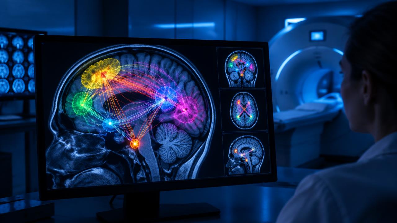
Brain Imaging Is Making TMS More Precise — and Possibly More Effective
Where exactly should the magnetic pulses land? For years, most TMS clinics have answered that question with a tape measure — using standardized scalp measurements to find the treatment target. Two randomized trials published in 2026 suggest that using each patient's own brain imaging may work better.
Connectivity-based targeting
A study published in JAMA Psychiatry randomized patients with depression to accelerated TMS aimed either by standard scalp measurements or by functional connectivity — maps showing how each person's prefrontal cortex connects to deeper mood-regulating regions. The connectivity-guided group improved significantly more, with a median depression score reduction of 24 points versus 18 points on the MADRS scale.
Individualized targets in a sham-controlled trial
A second trial, published in BMC Medicine, targeted TMS based on each patient's functional connectivity between the dorsolateral prefrontal cortex and the subgenual anterior cingulate — a hub implicated in depression. The individualized group saw a mean 64.3% reduction in HAMD-17 depression scores, compared with 41.4% in the sham group.
What this means for patients
Personalized targeting typically requires an MRI scan before treatment, which adds cost and is not yet standard everywhere. But the direction of the evidence is clear: depression is not identical from brain to brain, and treatment that accounts for those differences may work better. Expect "image-guided" or "neuronavigated" TMS to become a bigger part of the conversation with providers.
Sources
Written by
TMS News Editorial Team
The TMS News editorial team covers transcranial magnetic stimulation research, treatment access, and patient education, drawing on peer-reviewed studies and leading clinical centers.
 PreviousMeasuring TMS Progress: Why Response and Remission Are Different
 NextMotor Threshold in TMS: A Calibration Measure, Not a Depression Score